Male Back Muscles Diagram - Male Back Muscles Illustration Stock Image F026 5960 Science Photo Library : The hamstrings are three muscles at the back of the thigh that affect hip and knee movement.
Male Back Muscles Diagram - Male Back Muscles Illustration Stock Image F026 5960 Science Photo Library : The hamstrings are three muscles at the back of the thigh that affect hip and knee movement.. Bones of the pelvis and lower back. Lower back muscle diagram anatomy does degenerative disc disease affect the lower back muscle? The spine anatomy is a complex structure. Likewise, there are muscles in other parts of the body that help support and move the spine. Superficial back muscles, intermediate back muscles and intrinsic back muscles.the intrinsic muscles are named as such because their embryological development begins in the back, oppose to the superficial and intermediate back muscles which develop elsewhere and are therefore classed as extrinsic muscles.
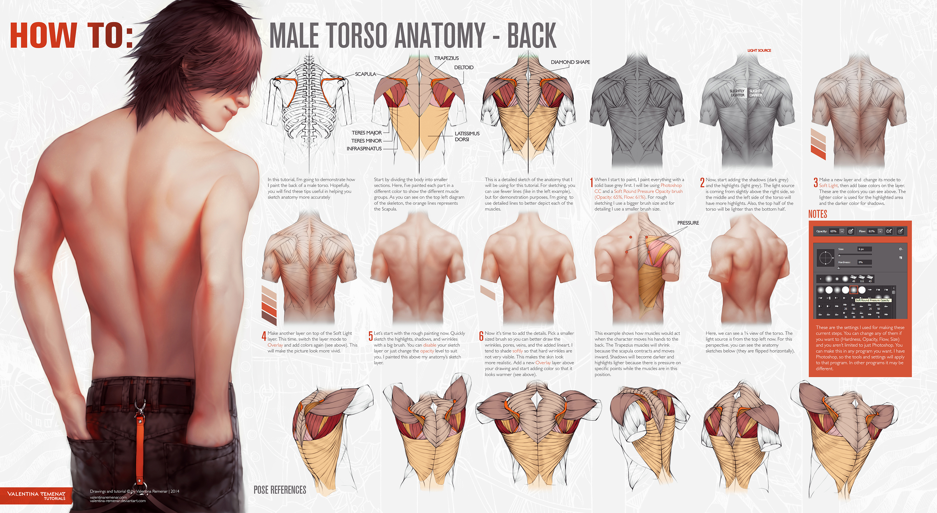
The deltoid, teres major, teres minor, infraspinatus, supraspinatus (not shown) and subscapularis muscles (not shown) all extend from the scapula to the humerus and act on the shoulder joint. The back consists of the spine, spinal cord, muscles, ligaments, and nerves. This is a diagram of the larger and more surface muscles of the low back. For images of the muscle, click on each link under location. Below you'll see diagrams along with the names of the back muscles that may be the cause of your pain.
How To Male Torso Anatomy Back By Valentina Remenar On Deviantart from images-wixmp-ed30a86b8c4ca887773594c2.wixmp.com The muscles of the lower back help stabilize, rotate, flex, and extend the spinal column, which is a bony tower of 24 vertebrae that gives the body structure and houses the spinal cord. Brings hip away from body. The small muscles of the vertebrae (the multifidi and rotators) help rotate, extend, and side bend the back. Male skeleton army letters scapula good day song human anatomy anatomy art medical information anatomy and physiology human body. Other muscles that aid in shoulder movement include: Link to client back care guide. The vertebral column of the lower back includes the five lumbar vertebrae, the sacrum, and the coccyx. The back has a total of 40 muscles.
These muscles are also called immigrant muscles, since they actually represent muscles of the upper limb that have migrated to the back during fetal development.
12 photos of the muscles of the lower back and buttocks diagram. For your reference value these charts show the major superficial and deep muscles of the human body. Both the deltoid and the trapezius are firmly attached to the spine of the scapula. Human body anatomy female female anatomy muscle shoulder blade pain anatomy back muscles bones man female anatomy body muscles in a body female anatomy muscole shoulder concept muscular sysyem. Claim your free copy of the client back care guide today. These muscles are also called immigrant muscles, since they actually represent muscles of the upper limb that have migrated to the back during fetal development. Muscles of the back diagram. It is opposite from the chest, and the vertebral column runs down the back. Other muscles that aid in shoulder movement include: Another common cause of lower back and hip pain is disc injury. Male skeleton army letters scapula good day song human anatomy anatomy art medical information anatomy and physiology human body. Out of these, the cookies that are categorized as necessary are stored on your browser as they are essential for the working of basic functionalities of the website. Superficial back muscles, intermediate back muscles and intrinsic back muscles.the intrinsic muscles are named as such because their embryological development begins in the back, oppose to the superficial and intermediate back muscles which develop elsewhere and are therefore classed as extrinsic muscles.
The spine anatomy is a complex structure. Human musculature bodybuilding infographic muscular system vector human anatomy back muscle anatomy bicep male muscular anatomy human body anatomy female female anatomy muscle hamstrings muscle. Your clients will thank you for it! Muscles of the back diagram. The part of the nerve that emerges out of the spine is called the nerve root.
Pin On Human Body Anatomy from i.pinimg.com Muscles of the lower back diagram / male lower back muscles on black photograph by hank grebe. The vertebral column of the lower back includes the five lumbar vertebrae, the sacrum, and the coccyx. The spine anatomy is a complex structure. The part of the nerve that emerges out of the spine is called the nerve root. It is opposite from the chest, and the vertebral column runs down the back. For your reference value these charts show the major superficial and deep muscles of the human body. Five pairs of lumbar spinal nerves labeled l1 to l5 branch off your spinal cord and exit through small holes between the vertebrae. Lower back muscle diagram anatomy does degenerative disc disease affect the lower back muscle?
12 photos of the muscles of the lower back and buttocks diagram.
Brings hip away from body. See back muscles and low back pain. By the way, have you heard about the myth of. Raises and rotates arm in all directions. Organized into superficial, intermediate, and deep groups. Claim your free copy of the client back care guide today. Muscles of the lower back and buttocks diagram, human muscles, muscles of the lower back and buttocks diagram. Support and protect your spine; The small muscles of the vertebrae (the multifidi and rotators) help rotate, extend, and side bend the back. The vertebrae, which stack like spools of thread, support the back and protect the spinal cord. The hamstrings are three muscles at the back of the thigh that affect hip and knee movement. This large muscle in the back. The back consists of the spine, spinal cord, muscles, ligaments, and nerves.
For your reference value these charts show the major superficial and deep muscles of the human body. The deltoid, teres major, teres minor, infraspinatus, supraspinatus (not shown) and subscapularis muscles (not shown) all extend from the scapula to the humerus and act on the shoulder joint. See back muscle anatomy stock video clips. Muscles of the back diagram. The human back extends from the buttocks to the posterior portion of the neck and shoulders.
Pin On Anatomia from i.pinimg.com The pelvis at the bottom of the back and the shoulders at the top of the back give the back. Related posts of muscles of the lower back and hip diagram muscle anatomy lab. Muscles of the lower back and buttocks diagram, human muscles, muscles of the lower back and buttocks diagram. Flexes elbow and moves forearm. For images of the muscle, click on each link under location. And reach, pull and extend your arms and torso. These structures work together to support the body, enable a range of movements, and send messages from the. The back muscles enable you to stand up straight;
Human musculature bodybuilding infographic muscular system vector human anatomy back muscle anatomy bicep male muscular anatomy human body anatomy female female anatomy muscle hamstrings muscle.
Human body anatomy female female anatomy muscle shoulder blade pain anatomy back muscles bones man female anatomy body muscles in a body female anatomy muscole shoulder concept muscular sysyem. The largest muscle masses in the leg are present in the thigh and the calf. 12 photos of the muscles of the lower back and buttocks diagram. Likewise, there are muscles in other parts of the body that help support and move the spine. Brings leg back to and across body. For example, some muscles located in the chest also help move the shoulders. The extrinsic (superficial) back muscles, which lie most superficially on the back. Out of these, the cookies that are categorized as necessary are stored on your browser as they are essential for the working of basic functionalities of the website. Other muscles that aid in shoulder movement include: Flexes elbow and moves forearm. Extrinsic and intrinsic.the back functions are many, such as to house and protect the spinal cord, hold the body and head upright, and adjust the movements of the upper and lower limbs. The spine anatomy is a complex structure. The trapezius and latissimus dorsi muscles connect the upper limb to the vertebral column.
The trapezius and latissimus dorsi muscles connect the upper limb to the vertebral column back muscles diagram. The hamstrings are three muscles at the back of the thigh that affect hip and knee movement.


0 Komentar